Cones (eye): important for color vision
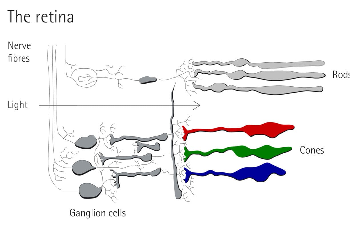
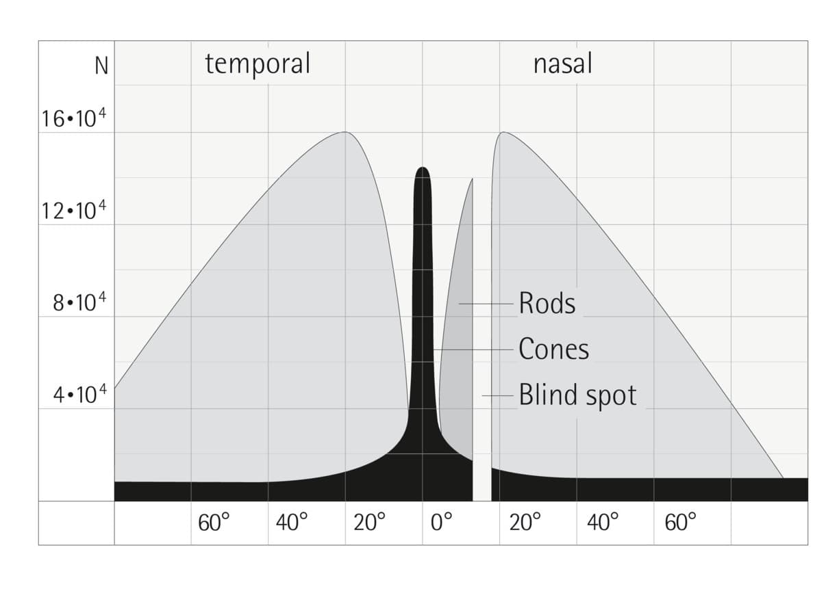
The cones are light-sensing cells in the eye that, with the rods, enable us to see. The cones in the eye determine our vision at higher luminous intensities, i.e. during the day or with artificial lighting. The system of cones has low sensitivity to light and is mainly concentrated in the central area around the fovea. It enables color vision and very good visual acuity.


What tasks do cones in the eye have?
Cones are photoreceptors, meaning specialized light-sensing cells of the retina in the eye. The cones enable vision in sufficient light, i.e. mainly during the day and in artificial lighting. They are vital for the perception of colors.
The retina of the eye has around six million cones, i.e. less than rods. But it is only through these cone-like receptors that the eye is capable of visual acuity. The concentration of cones is highest in the fovea (fovea centralis), the retinal area with the sharpest vision.

What are L, M and S cones?
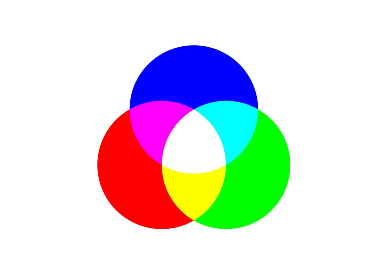
There are three different types of cones in the human eye. They are distinguished on the basis of their spectral absorption curve. The focus of sensitivity is in the blue, green and red spectral range. The balanced stimulus response of the three cone types forms a white color impression and is the basis of colorimetry.
- S cones (short wavelength receptor): This receptor covers the blue range of the spectrum visible to humans and is therefore also called a blue receptor. They are the least common.
- M cones (medium wavelength receptor): M cones are also called green receptors and mainly cover the green range, i.e. between blue and orange light. These receptors are the second most common in the retina of the eye.
- L cones (long wavelength receptor): L cones have maximum perception in the long-wave range. They are also called red receptors, because with yellow-green they contribute significantly to the perception of this color. In most people, L cones are the most common.


